
Dawson's Sore Mouth
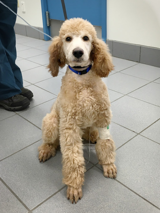
This is Dawson! Dawson is a goofy, sweet, perpetual motion machine who came into the clinic recently for an examination and vaccines. This is probably one of very few photos of Dawson sitting still - there were about 50 photos I had to comb through to find one that wasn't a blonde blur.
During Dawson's exam, his vet noticed that the deciduous (or baby) canine teeth on his bottom had not come out in the correct position, and were digging into the soft tissue of his hard palate. This is termed "lingual displacement of mandibular canine teeth" or "base narrow canines," and is something that we routinely check for when examining puppies.
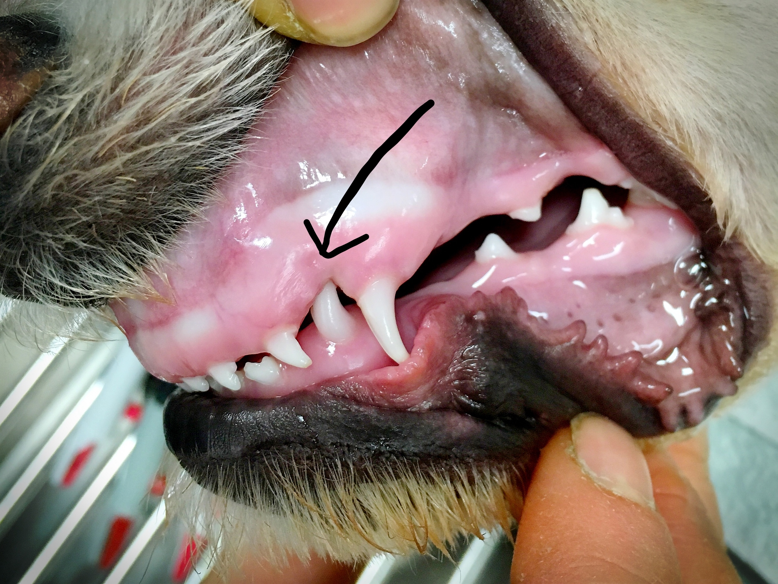
I take and edit all the photos for the blog on my phone, so my arrow drawings leave something to be desired. This arrow is pointing to the canine tooth on the bottom jaw embedded in the soft tissue of the upper jaw.

This picture is looking at Dawson's upper jaw, and the two arrows are pointing to the holes created by the canine teeth piercing through the soft tissue.
This is obviously a very painful condition for puppies, as every time they close their mouth their teeth are penetrating and traumatizing the sensitive soft tissue on their upper jaw. In addition to being painful, these permanent wounds act as a portal for infection, and puppies can develop infection in the bone of their upper jaw or in their nasal sinuses as a consequence of this condition.
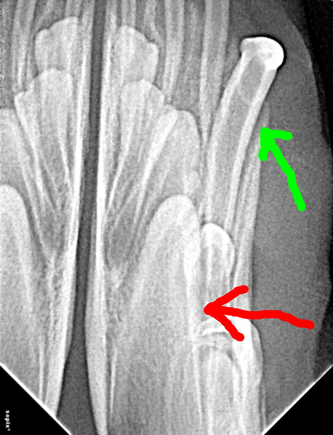
Treatment for this condition is removal of the misplaced teeth. Before beginning surgery, Dawson's vet took an x-ray of his bottom jaw to ensure that there weren't any surprises hiding, like a fracture of the canine tooth, infection, or loss of the root. The green arrow is pointing at the offending canine tooth, and the red arrow is pointing at the permanent canine tooth that will erupt in 1-2 months. I find x-rays of puppy and kitten mouths fascinating, because you can see the developing permanent teeth hidden in the bone like seeds waiting to sprout.
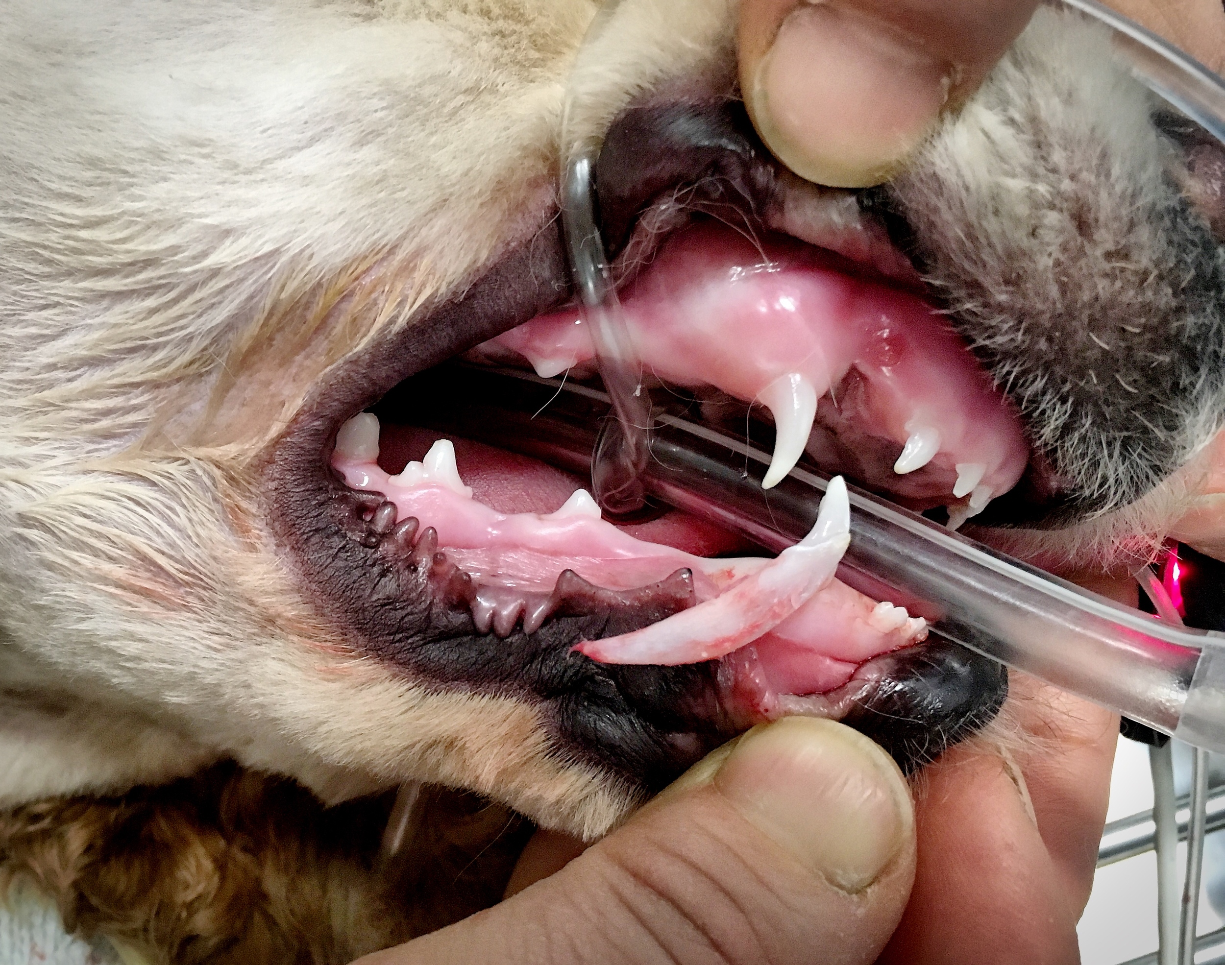
This next picture shows Dawson's extracted canine tooth next to its surgical site. The crown of the tooth, or the visible portion that sits above the gumline, is only about one third of the total length of the tooth - these teeth can be challenging to extract as they are very delicate with thin walls and long, tapering roots, and can break if extracted improperly.
Dawson's surgical sites are closed with absorbable suture, which will break down over the next 2-3 weeks. Both of Dawson's bottom canine teeth were affected and removed, but many puppies will be affected on only one side.

These surgeries are fun because they're technically challenging and provide instant gratification - as soon as the dog wakes up they're immediately more comfortable and happy. Because these were the deciduous or baby teeth, we will have to wait and see how Dawson's permanent canine teeth erupt. In many cases the permanent teeth will erupt in the correct position, but if Dawson's permanent teeth are also improperly positioned then we will have to discuss the different treatment options available for managing the permanent teeth.