
Charlie's Mast Cell Tumour
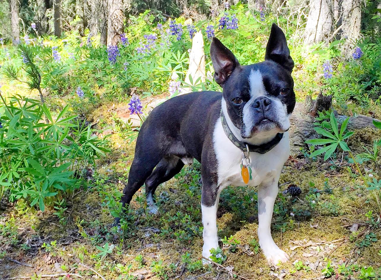
This is Charlie! Charlie is an irresistible Boston Terrier whose favourite things include food, staring at you without blinking if you're eating food and not sharing, sleeping under the covers, his little spider toy, and his family. Several years ago Charlie had a mass removed from his hind end that was diagnosed as a mast cell tumor, so when he developed another mass between his shoulder blades, his owners brought Charlie in immediately so we could see if he had another mast cell tumour.
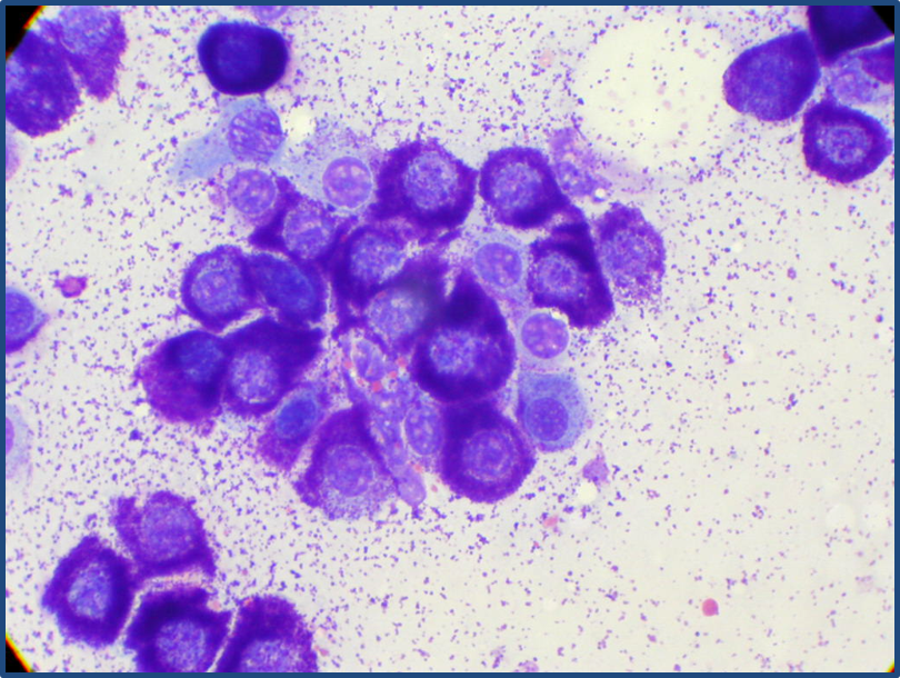
These are cells from a mast cell tumour viewed under a microscope. The dark purple cells have a high density of granules, and the purple speckles are free granules that have been released from mast cells.
Mast cell tumours are one of the most common types of of tumours that we diagnose in dogs. Mast cells are a type of white blood cell that play an important role in allergic responses. They have characteristic dark purple staining granules, making them usually easy to identify under the microscope. Mast cell tumours found in the mouth, on the muzzle, or around the genitals tend to behave more aggressively than mast cell tumours found on other parts of the body. Some mast cell tumours can behave aggressively and spread to other locations in the body, such as lymph nodes or internal organs. Even if a mast cell tumour does not spread to other locations, it can still cause disease through release of histamine, which increases stomach acid production and can result in stomach ulcers. Boxers and Boston Terriers seem to be more susceptible to mast cell tumours than other dog breeds.
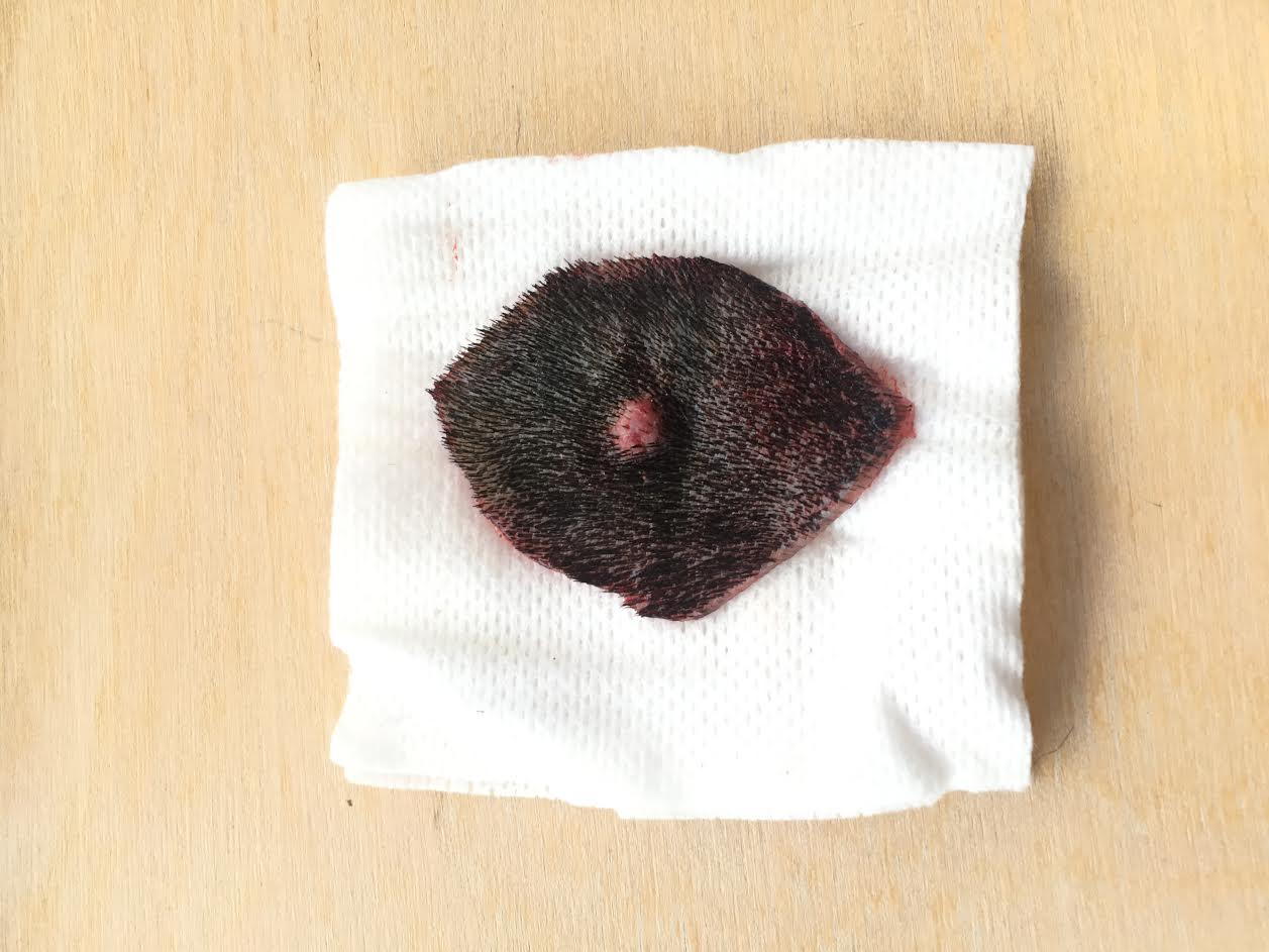
Charlie's mast cell tumour.
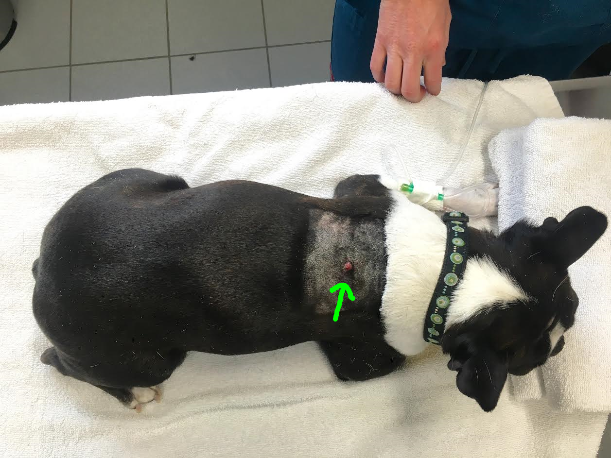
Surgery is the treatment of choice for mast cell tumours, so we were very lucky that Charlie's tumour was on his back and not in a difficult location, such as near an eye or on the muzzle. When surgically removing any tumours, we have to remove margins of normal looking tissue around the tumour to ensure that there are no tumour cells left behind. This pre-surgical picture of Charlie shows how we were planning on removing his tumour - the green circle shows how much normal-looking tissue around the tumor has to be removed. The following picture shows Charlie's tumour after removal.

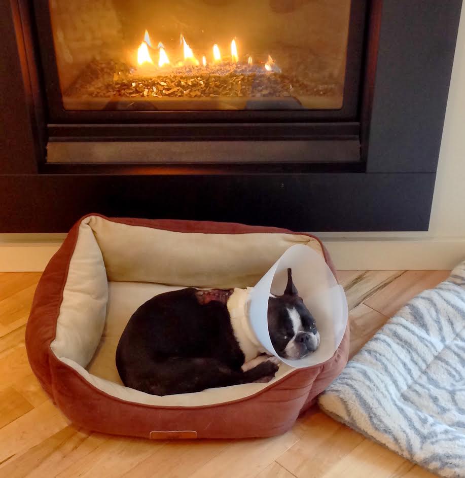
Charlie's surgery went very well and he's recovering at home (quite stylishly in front of the fireplace). Charlie's tumour has been sent down to the pathology lab in Vancouver to determine what how aggressive the tumour is and whether or not Charlie will require any followup treatment. The pathologist will also examine the surgical margins and let us know how likely it is that Charlie's surgical site is contaminated with tumour cells. Because Charlie is a Boston Terrier, he may unfortunately develop another mast cell tumour in his lifetime, but he's lucky to have an amazing family that is keeping a close eye on him and monitoring him for any new masses.